Hepatocytes directly isolated from liver tissue show a functionality very similar in culture as in vivo. They have become the “gold standard” for metabolism, clearance, hepatoxicity and drug-drug interaction and a lot more. The development of various mouse models have increased the availability of tissue samples over the past two decades.
Isolating hepatocytes from liver tissue samples is either time-consuming, non-standardized, not scalable or accepting all drawbacks of enzymatic digestion. Or all of them together. This is all the more important because it also implies major limitations for a 2D or 3D culture.
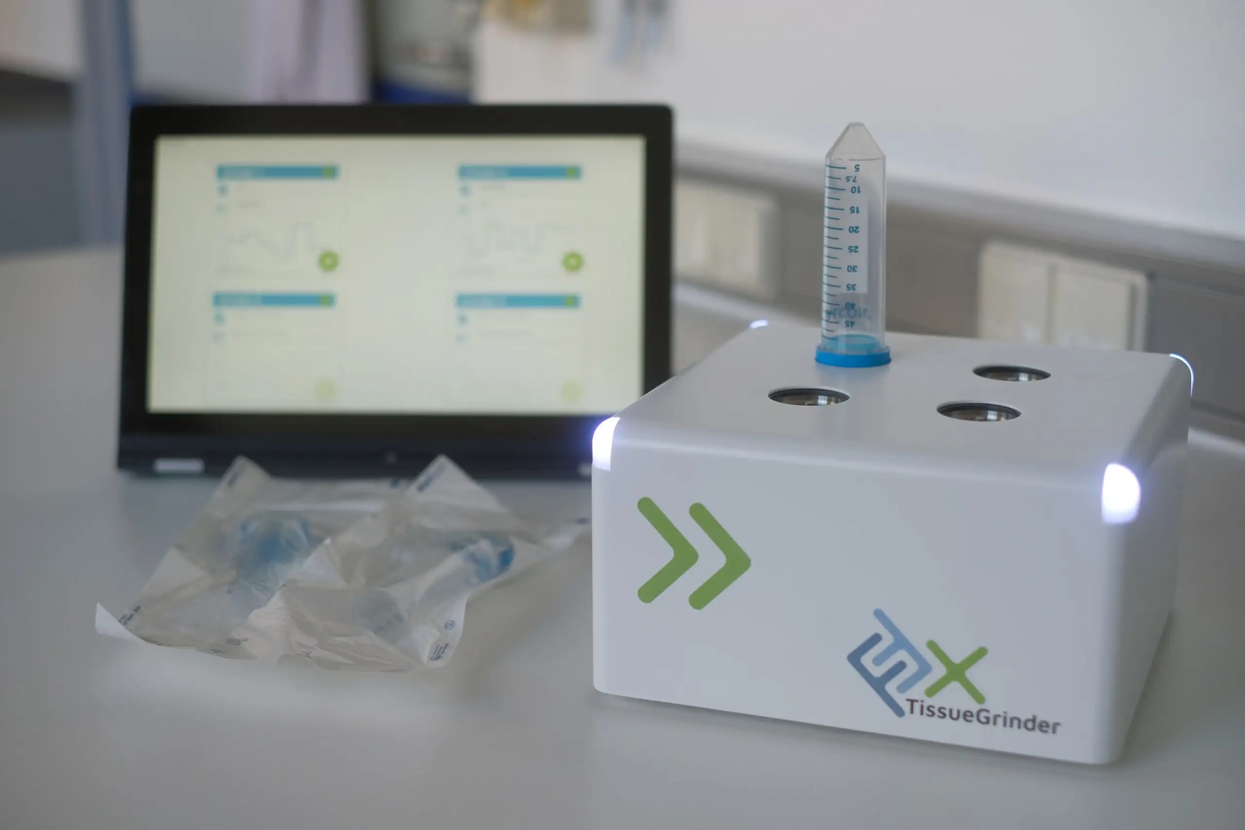
With the FFX TissueGrinder, liver samples can now be dissociated into single cell suspensions within a matter of just a few minutes enzyme-free. It is therefore a superior new option for obtaining a high yield of premium viable cells maintaining surface markers.
Advantages of Primary Hepatocyte Isolation Without Enzymes
To obtain primary hepatocytes from liver samples, the FFX TissueGrinder offers great advantages:
Due to the enzyme-free approach, processing time is significantly reduced and
the properties of the hepatocytes are preserved,
not to mention that some enzymatic methods do not yield hepatocytes at all
The contamination of the sample is effectivly prevented by the integration of the grinding component together with a cell strainer into a centrifugation tube.
This improves the quality of any subsequent cell culture significantly.
Applications of Primary Hepatocyte Isolation in Research
In a flow cytometry scatterplot, hepatocytes are easily visible, when the tissue sample is dissociated with the TissueGrinder.
In contrast, hepatocytes cannot be analyzed with enzymatic tissue dissociation.
The TissueGrinder gives a good yield of hepatocytes, while there are close to none retrievable from the enzymatic process.
The generated primary cells can be employed to create tissue models such as spheroids, micro tissues, organoids and cell printed systems. New cell lines can easily be established for molecular cell characterization and drug screenings.
enzymatic digestion
TissueGrinder dissociation
Day 0
d0
Day 1
d1
Sounds too good to be true?
Then contact us to arrange a test run with your own samples!