Single Cells from FFPE samples for Flow Cytometry and NGS

Next Generation Single-Cell Sequencing (NGS) is becoming a valuable standard method in life science research and personalized medicine.

Formalin-fixed paraffin-embedded (FFPE) tissue samples are a relevant source of material for this because of its wide availability and broad spectrum of sample types.

However, making high quality single cells from FFPE samples accessible for NGS is a challenge. Reflecting the tumor or tissue heterogeneity to uncover different genetic and phenotypic characteristics is a real bottleneck.

 
Diagram showing the workflow of single-cell sequencing, from cell isolation to DNA/RNA extraction, amplification, and genomics analysis.

Tang et al. Cell Biosci. (2019) 9:53

Sample Input

Sample Input

FFPE 50 µm sections*

Sample Preparation

  • Deparaffinization
  • Rehydration
  • Antigen Retrieval
Preparation
Enzymatic

Enzymatic Pretreatment

Minimized 20 min enzyme treatment

Dissociation

Fast but gentle mechanical dissociation with the TissueGrinder

TissueGrinder
Staining

Cell Staining

Antibody labeling
Cytospins

Cell Sorting

FACS Isolation of pure tumor and stromal cell populations

Sorting
Lysis

Cell Lysis

Access to pure tumor and stromal cell DNA

NGS and Data Analysis

Library preparation & detection of tumor specific characteristics

NGS
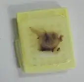
We established a protocol* for generating relevant material of single cells from FFPE samples in reasonable time to support subsequent analysis.
 
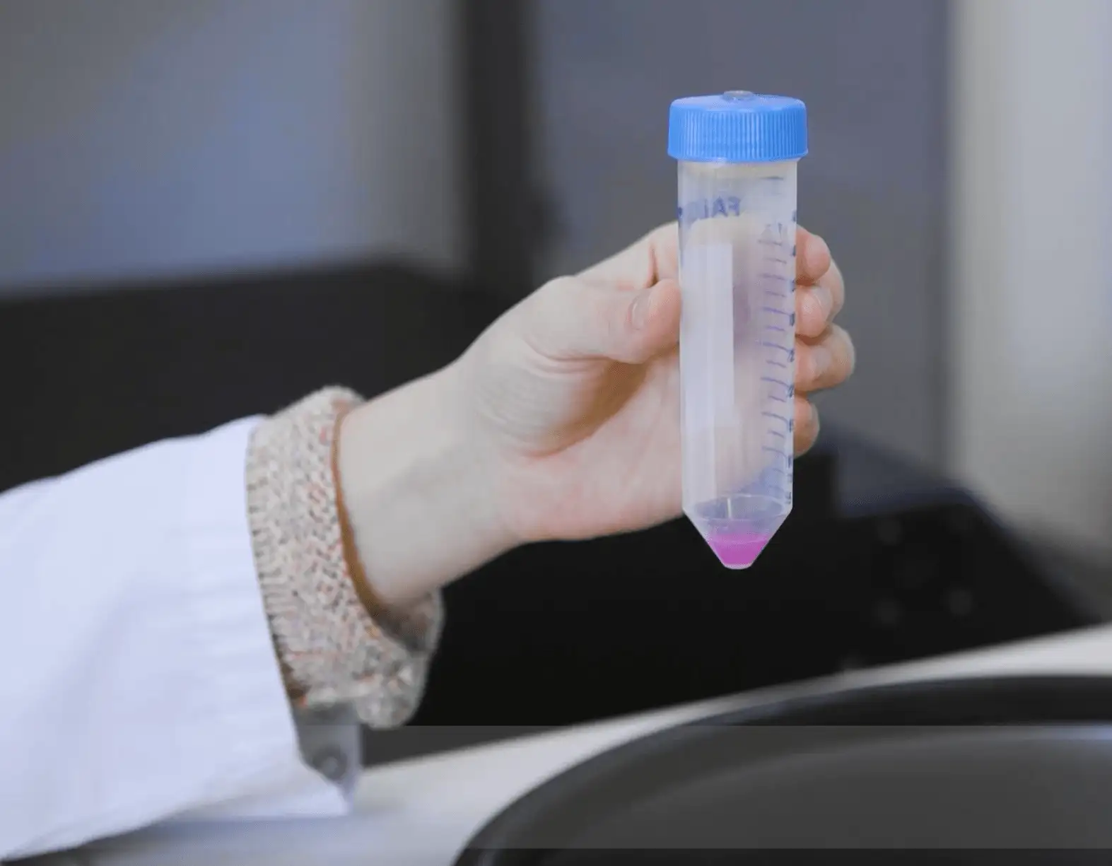
After removing excess parafin, the tissue is cut into 4 sub-samples with 100-200mg and transferred into an Eppendorf Cap.
 
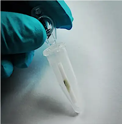
The samples are then incubated in a water bath at 65°C for 25 min using HistoChoice as a xylene substitute.
 
By incubating in a decreasing ethanol series for 2 x 15 min, the sample is rehydrated.
 
With repeated washing in ARS and DPBS, the masked epitopes in the tissue are unmasked and lost immunoreactivity is partially restored within 60 min.
 
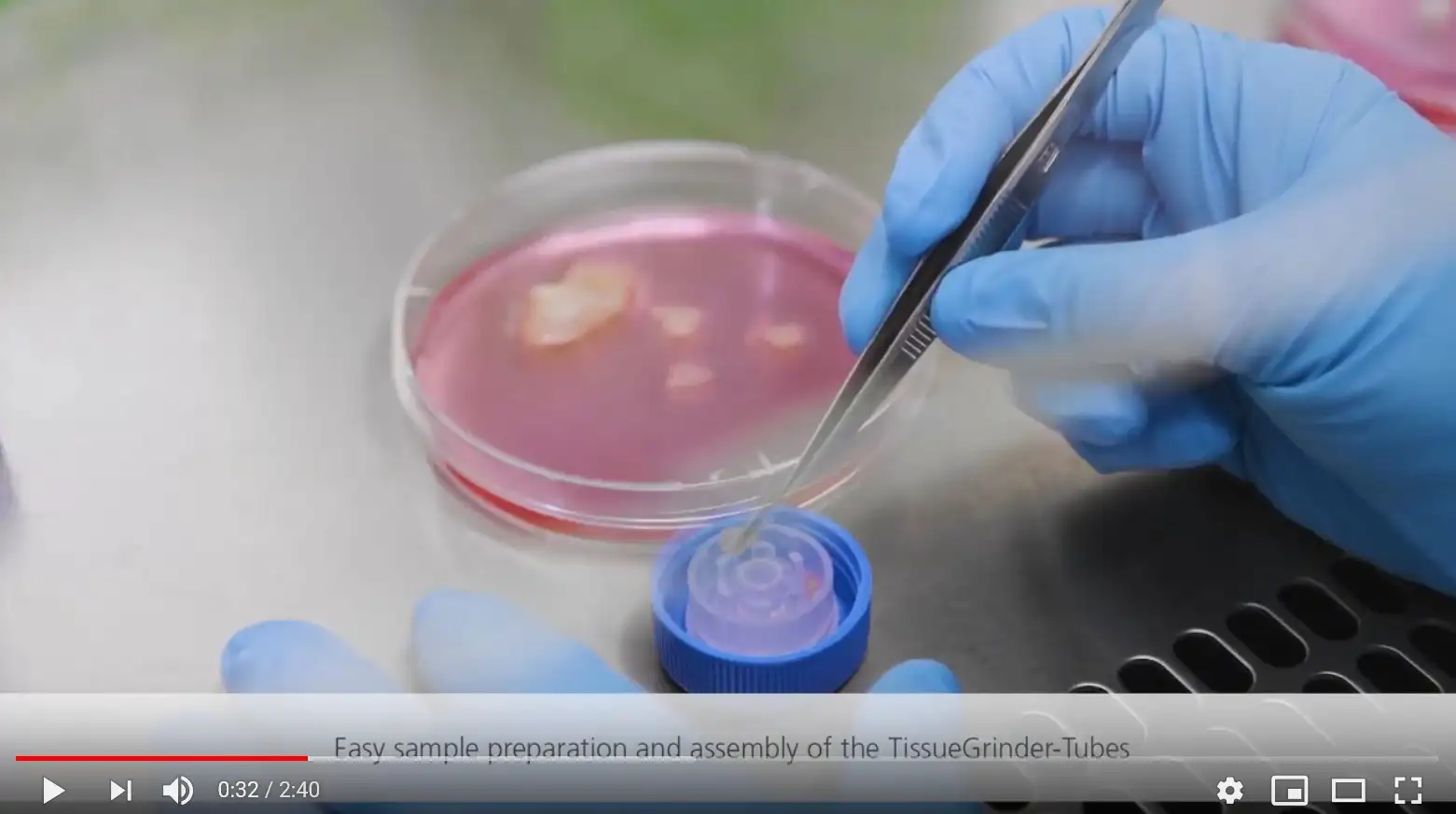
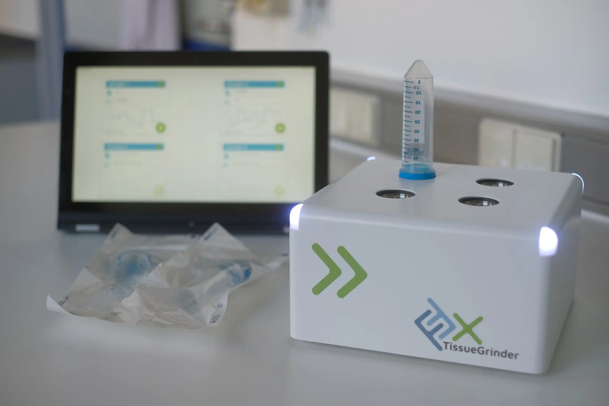
With a minimal enzymatic pre-treatment, the subsequent mechanical dissociation with our TissueGrinder makes the cells from the tissue accessible to immunofluorescent staining, flow sorting and subsequent molecular analyses by e.g. next generation sequencing.
 
The enzymatic incubation time can be reduced from 50 to 20 minutes, resulting in faster workflow and much higher quality of the cells obtained.

* TissueGrinder optimised FFPE protocol is based on the following study: https://link.springer.com/article/10.1186/s10020-019-0108-z

The 40x microscopy image show examples of how the single cells from FFPE samples generated with our protocol are suitable for immunofluorescent staining.

Keratin

Vimentin

DAPI

Immunofluorescence image of cultured cells stained with blue nuclear, green cytoskeletal, and red functional markers on black background.
Fluorescence microscopy image of cells stained with green cytoskeletal, blue nuclear, and red functional markers showing diverse cell shapes and expression patterns.
Fluorescence microscopy image of adherent cells stained with green for cytoskeleton, blue for nuclei, and red for additional markers.
Immunofluorescence image showing single cells stained with green for cytoskeleton, blue for nuclei, and red for functional markers on black background.

The single cells obtained with the TissueGrinder process are in a much better condition than those produced with a conventional enzymatic automation.

Fluorescence microscopy image showing stained single cells with blue nuclei and green membrane markers on a black background.

On the left, an example of single cells generated from FFPE samples with the TissueGrinder compared to a standard enzymatic dissociation on the right.

Above, an example of single cells generated from FFPE samples with the TissueGrinder compared to a standard enzymatic dissociation below.

Fluorescence microscopy image showing tissue-derived cells stained with blue, green, and red fluorescent markers for multi-parameter analysis.
Close-up of TissueGrinder tube components with transparent grinding inserts and blue cap for tissue dissociation.
TissueGrinder benchtop device for automated mechanical tissue dissociation in laboratory workflows.
Hand holding a 50 ml centrifuge tube with blue cap and pink solution for tissue processing.

Sounds too good to be true?

Then contact us to arrange a test run with your own samples!