Surface markers are essential biomarkers for precise diagnosis, disease monitoring, and therapy prediction. Inadequate marker expression can lead to misdiagnoses and inappropriate treatments. Thus, selecting the right dissociation technique for single-cell isolation is crucial to preserving the integrity of these markers.
Splenocytes were incubated with collagenase IV, V and dispase (0.5 U/ml) for different time periods. The prevalence of markers is presented as normalized means ±SD (n=3) relative to the initial incubation time.
Splenocytes were incubated with various concentrations (0.25–1.0 U/ml) of collagenase IV, V and dispase for 10 minutes. The prevalence is shown as normalized means ±SD (n=4) relative to the initial enzyme concentration of 0 U/ml.
Enzymatic treatment of splenocytes resulted in a significantly reduced surface marker expression of CD27 and CD62L with increasing incubation time and enzyme concentration of collagenase IV, V, and dispase.
Comparing enzymatic with mechanical tissue dissociation
To facilitate the isolation of single cells, semi-automated methods, gentleMACS (enzymatic and mechanical) and TissueGrinder (purely mechanical), were compared for single-cell isolation. TissueGrinder’s non-enzymatic approach demonstrated faster and gentler dissociation while better preserving surface markers like CD27 and CD62L, resulting in a more representative cell suspension reflecting the actual cell population and morphology in the tissue, also facilitating better surface marker detection.
Monitoring the immunization process and the activation of leukocytes
Accurate expression of cell surface markers is essential for monitoring the immunization process and the activation of leukocytes during vaccine development. Vector-based vaccines deliver target antigens into the host to induce a specific immune response. However, faulty marker expression can lead to inaccurate results when assessing vaccine efficacy, as it may misrepresent immune cell activation and give a false impression of the vaccine’s effectiveness.
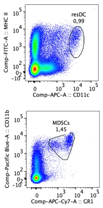
Manual with enzymes
Conversely, mechanical dissociation with the TissueGrinder provides superior preservation of dendritic and myeloid suppressor cells compared to enzymatic methods, which can compromise cell morphology. Accurately differentiating between various cell populations is crucial, as each fulfills distinct roles in the immune response.
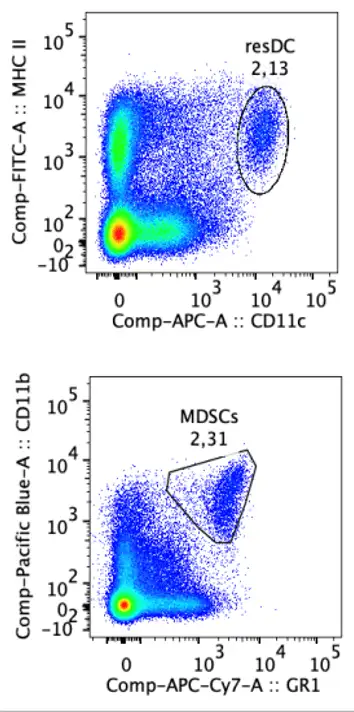
TissueGrinder
Conversely, mechanical dissociation with the TissueGrinder provides superior preservation of dendritic and myeloid suppressor cells compared to enzymatic methods, which can compromise cell morphology. Accurately differentiating between various cell populations is crucial, as each fulfills distinct roles in the immune response.
Manual with enzymes
TissueGrinder
Mice immunized with OVA-vectors exhibited activation of cytotoxic T cells, with concurrent expression of CD43 and CD27 signifying a strong immune response essential for the effectiveness of vector vaccines. However, enzymatic treatments may damage surface markers like CD27, leading to inaccurate assessments of cell activation and vaccine efficacy.
Sounds beneficial for your application?
Then contact us to arrange a test run with the TissueGrinder for your own samples and improve your surface marker detection!