Mechanical tissue dissociation enables the generation of patient-derived microtumors and tissue-derived microtissues that preserve cellular heterogeneity and support reliable functional drug screening.
Generating biologically relevant 3D tumor models from patient tissue remains a major challenge in translational research. In contrast to enzymatic digestion, mechanical dissociation helps preserve the viability of fragile primary cell populations and ECM-associated components, providing a more suitable starting point for functional assays and advanced 3D model workflows.
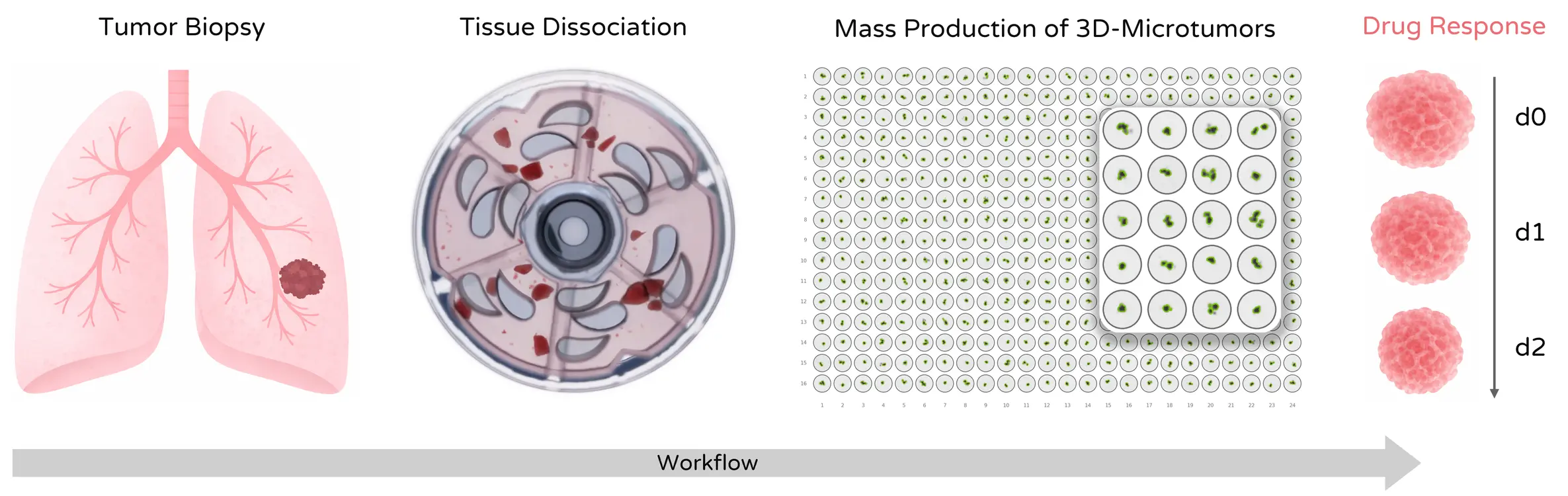
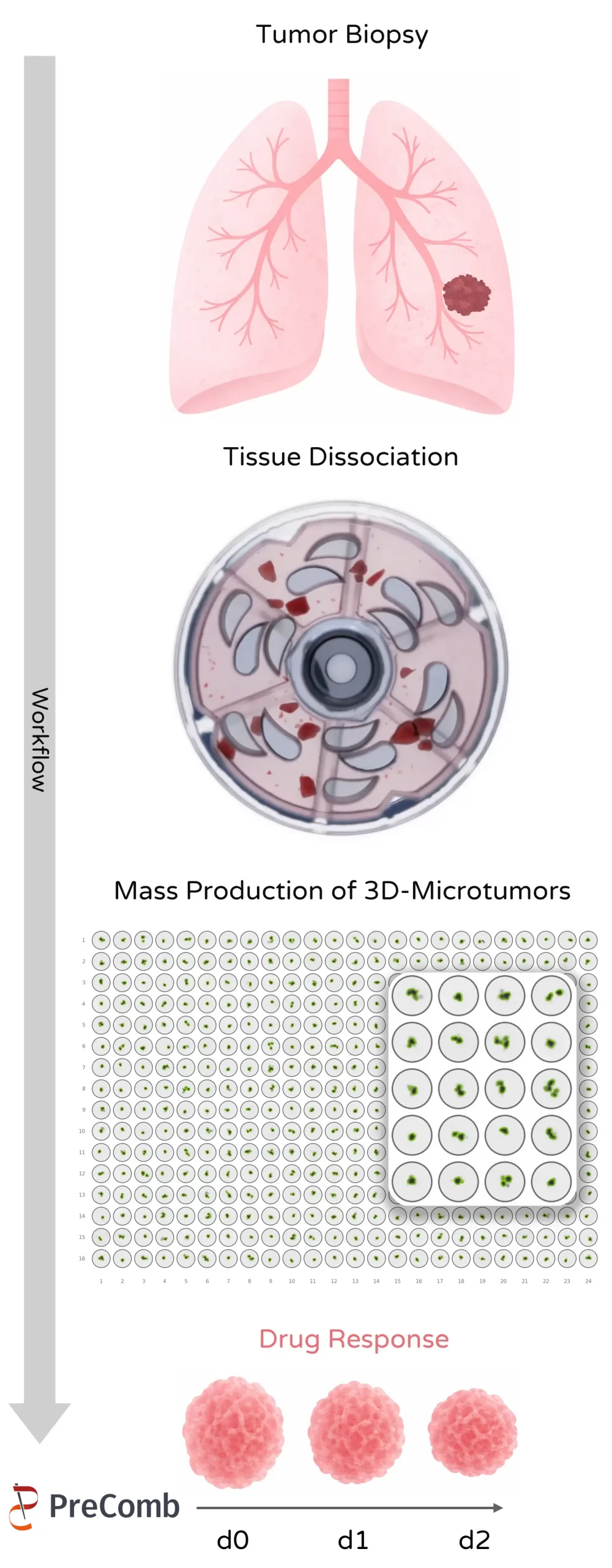
Mechanical processing offers a gentle and efficient alternative approach that maintains cell viability and supports the formation of functional microtumor systems.
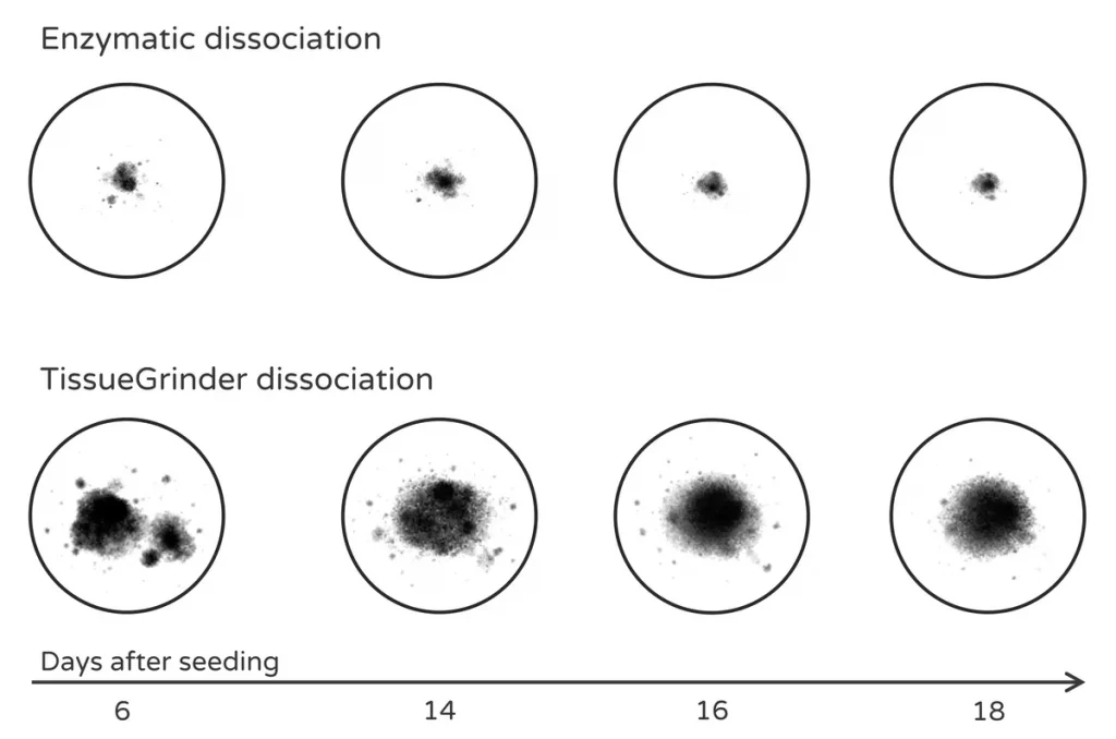
Recent work demonstrates that mechanical dissociation supports the formation of larger, more stable microtumors compared to enzymatic processing, supporting long-term functional assays and drug response studies.
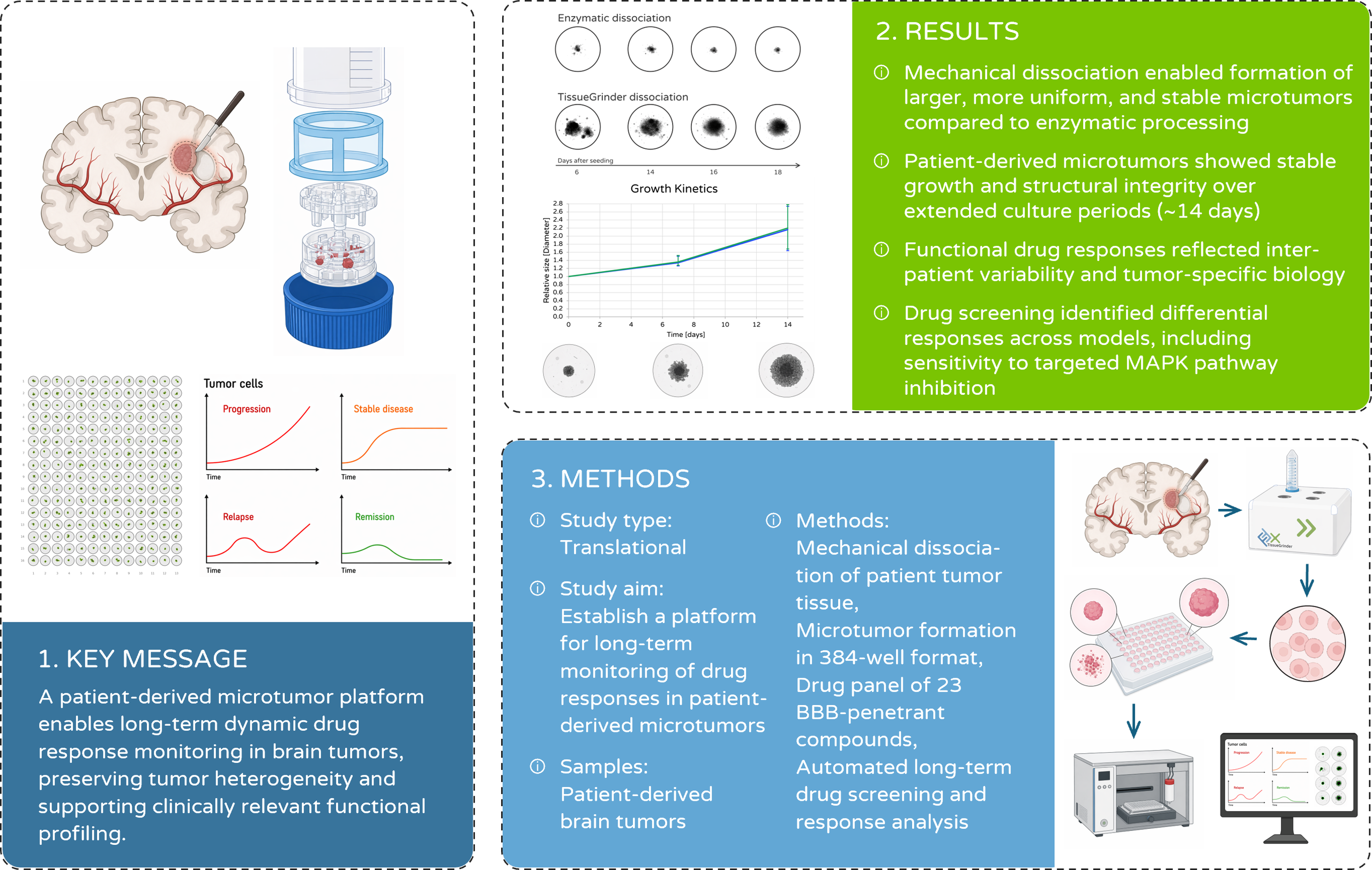
Reference: M. Bouzereau, M. Freitas, J. Kelm, S. Manser, E. Le Rhun, M. Weller, T. Weiss (2025) – Long-term functional drug and immunotherapy screening in immune-competent patient-derived microtissues across brain tumors, Neuro-Oncologys V27, Supplement 5 doi:10.1093/neuronc/noaf201.137
Preservation of viable primary cell populations:
Instead of extensive enzymatic digestion, mechanical dissociation helps preserve fragile primary cells and ECM-associated components as a suitable starting point for downstream microtumor formation.
Formation of stable and uniform microtumors and tissue-derived microtissues:
This results in microtumor structures that form reliably after seeding and show consistent size and morphology across experiments.
Reduced dependency on external stromal support (e.g. astrocytes):
Microtumors can form directly from the patient sample without requiring additional supporting cell types to stabilize the system.
Improved reproducibility compared to enzymatic methods:
By avoiding enzyme-related variability, results become less dependent on timing and batch-to-batch differences.
Compatibility with high-throughput screening platforms:
The workflow integrates naturally into standard plate-based formats, making it suitable for parallelized drug testing.
Maintained cellular heterogeneity and functional relevance:
Viable and diverse tumor cell populations enable more meaningful and representative functional readouts.
Fast and scalable sample processing:
Mechanical dissociation enables rapid and straightforward tissue processing, reducing handling complexity and enabling scalable workflows for larger sample numbers compared to enzymatic digestion.
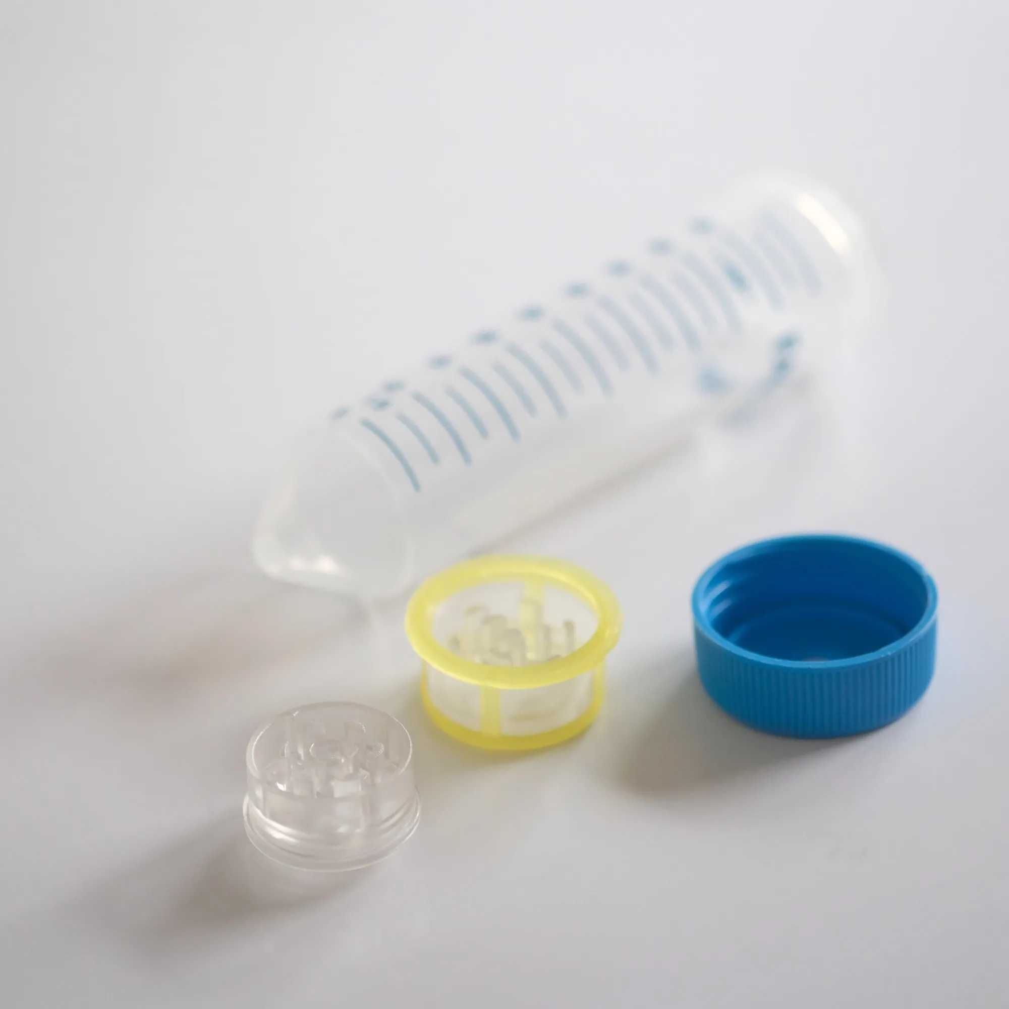
From Tissue to Functional Microtumor Assay
3DTwin® Screening method established at PreComb
Automated Mechanical Dissociation enables the generation of: