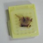
FFPE 50 µm sections*
Deparaffinization
Rehydration
Antigen Retrieval
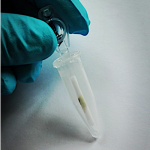
Minimized 20 min enzyme treatment
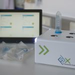
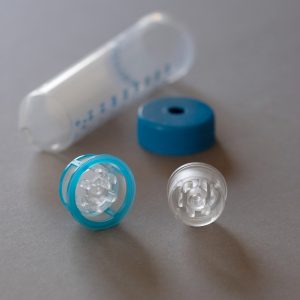
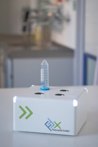
Fast but gentle mechanical dissociation
with the TissueGrinder
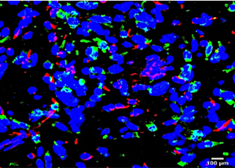
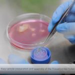
Antibody labeling
Cytospins
FACS
Isolation of pure tumor and stromal
cell populations
Access to pure tumor and stromal cell DNA

Library preparation
Detection of tumor specific genetic characteristics on cell populations